Home

ביקורת שלנו מצורף posterior pole of eye ל לזהם אהוב

Middle-aged man reports new onset diplopia

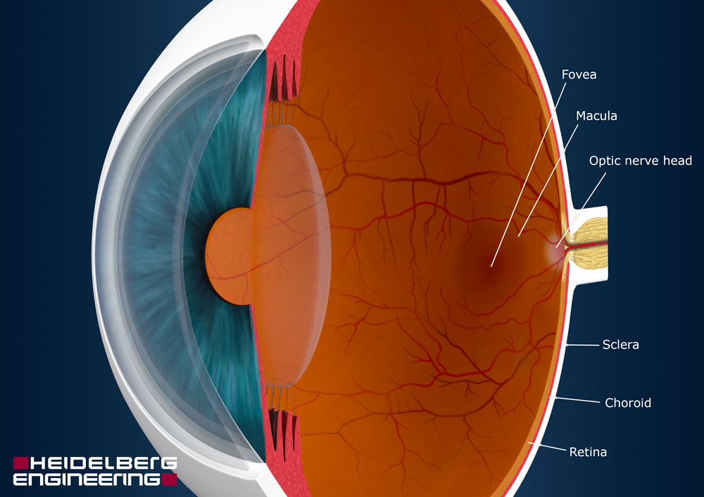
Retina: Anatomy, Function, and Treatment

TheEye
TheEye

Anatomy Of Retina - www.medicoapps.org

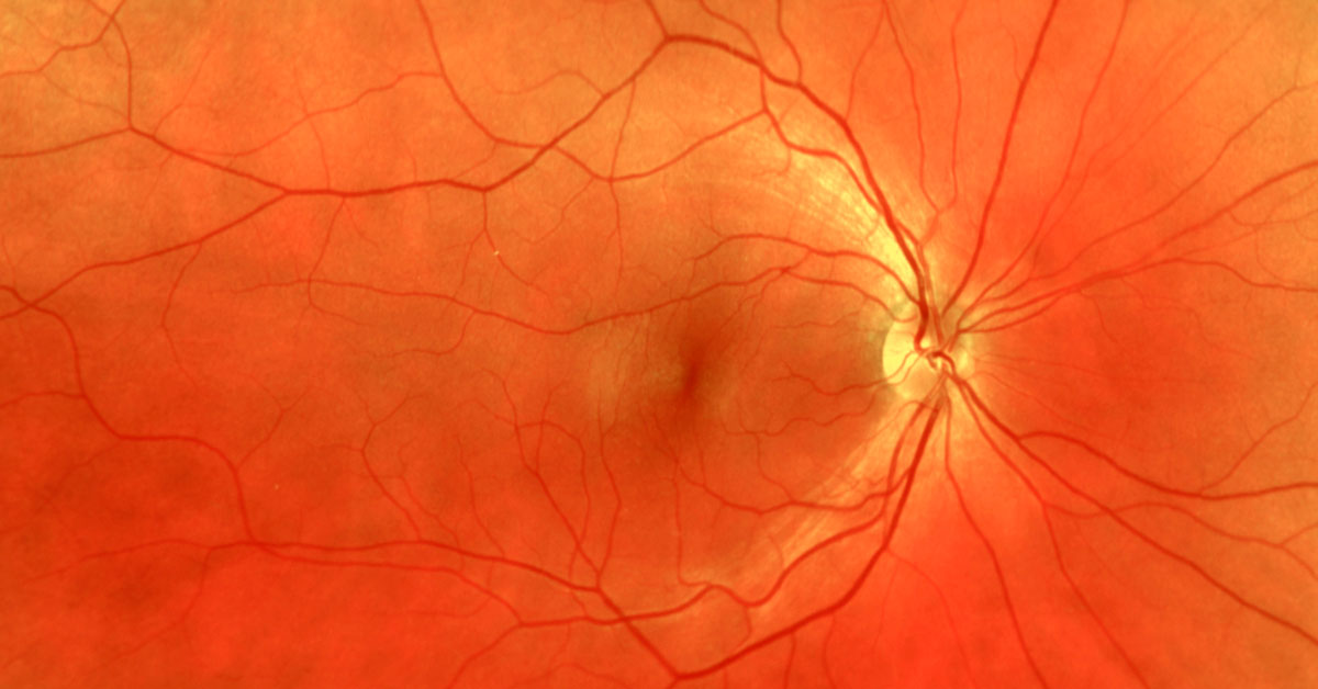
Posterior pole of eyeball - e-Anatomy - IMAIOS

Fundus images of the posterior pole of the right (A) and left (B) eyes... |  Download Scientific Diagram
Fundus images of the posterior pole of the right (A) and left (B) eyes... | Download Scientific Diagram

A) The posterior pole of the right eye shows multiple folds and an... |  Download Scientific Diagram
A) The posterior pole of the right eye shows multiple folds and an... | Download Scientific Diagram

Human eye - Extraocular Muscles | Britannica

Acute lymphocytic leukemia relapsing as bilateral serous retinal  detachment: a case report | Eye
Acute lymphocytic leukemia relapsing as bilateral serous retinal detachment: a case report | Eye

Posterior pole of eyeball - e-Anatomy - IMAIOS

▷ What is the POSTERIOR POLE OF THE EYE? - Área Oftalmológica

Posterior Pole and Peripheral Retinal Fibrovascular Proliferation in von  Hippel‐Lindau Disease | Semantic Scholar
Posterior Pole and Peripheral Retinal Fibrovascular Proliferation in von Hippel‐Lindau Disease | Semantic Scholar

Posterior pole of the retina, eye schematic - #AN0009

The yellowish pigmented spot at the posterior pole of the human eye lateral  to the blind spot isA. CristaB. SacculeC. IrisD. Macula luteaE. Meatus
The yellowish pigmented spot at the posterior pole of the human eye lateral to the blind spot isA. CristaB. SacculeC. IrisD. Macula luteaE. Meatus

JaypeeDigital | eBook Reader

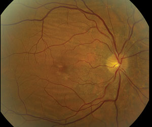
Fundoscopic photograph of a normal retina indicating the regions:... |  Download Scientific Diagram
Fundoscopic photograph of a normal retina indicating the regions:... | Download Scientific Diagram

Functions and pathologies of the posterior segment of the eye

Sensory Organs: Eye and Ear Flashcards | Quizlet

A Wrinkle, in Time
A Wrinkle, in Time

Eyeball (Bulbus Oculi) – Earth's Lab

Internal Structures of the Eye Diagram | Quizlet

Point at posterior pole, which is jmedial to and slightly above erve leave  is - YouTube
Point at posterior pole, which is jmedial to and slightly above erve leave is - YouTube

Gross Anatomy of the Eye Cornea at anterior –Light passes to lens Retina at  posterior –sensory tissue –sensory cells: rods and cones. - ppt download
Gross Anatomy of the Eye Cornea at anterior –Light passes to lens Retina at posterior –sensory tissue –sensory cells: rods and cones. - ppt download

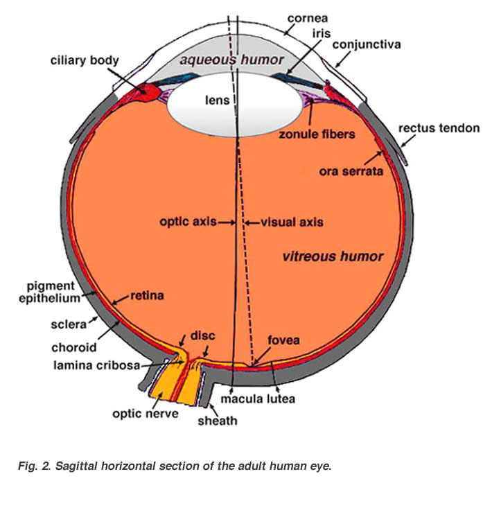
Gross Anatomy of the Eye by Helga Kolb – Webvision

Posterior part of your eye

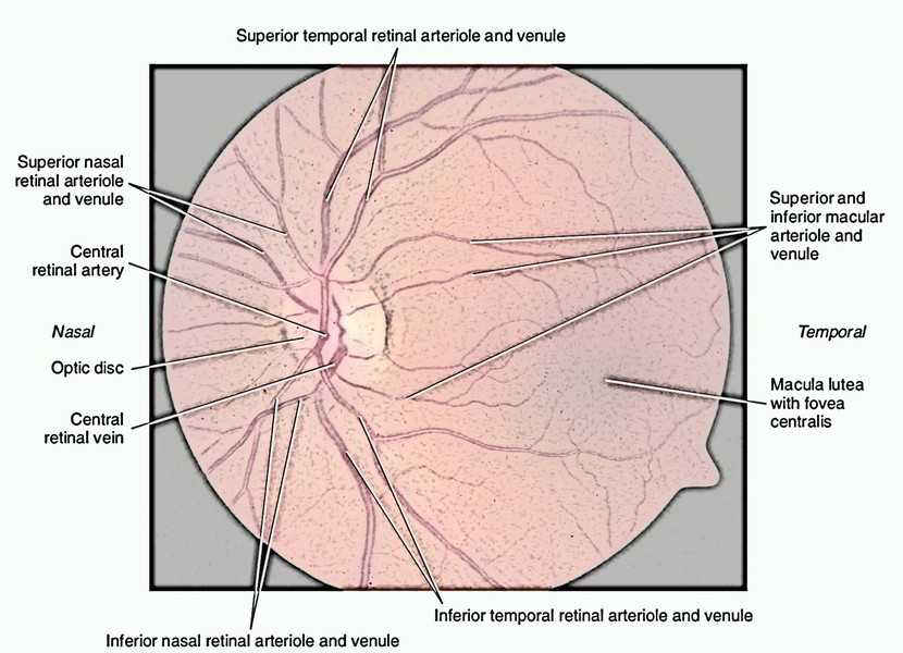
Anatomy of Retina
Anatomy of Retina

Eyeball: Structure and function | Kenhub